Procedure
When the LTT for allergens is performed, mononuclear cells are isolated from the patient’s heparinised venous blood under sterile conditions using density gradient centrifugation. Following several washing steps, these cells are cultured together with the test allergens (e.g. medications, metals, environmental allergens, among others) under optimised cell culture conditions over a period of 5 days.
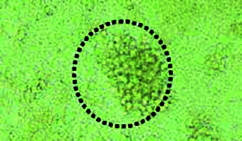
In patients with allergen-specific sensitisation, i.e. where lymphocytes specifically recognising the allergen circulate in the blood, these lymphocytes are activated to start clonal proliferation.
After day 5, 3H-thymidine is added to the cell culture for further 12 hours. This is integrated into the newly formed DNA of activated (dividing) lymphocytes.
Based on the content of 3H-thymidine-marked DNA, the cell division rate in the respective culture and thus the cell activation in this culture can be quantified.

To show the result, the allergen-induced lymphocyte proliferation is calculated in comparison with the spontaneous proliferation (blank) and the result is reported as stimulation index. The stimulation index is the ratio of the proliferation rates in the allergen-stimulated test run and the non-stimulated blank.
If the LTT is used to detect allergic sensitisations, SI values below 2 are regarded as negative (no sensitisation).
Results between 2 and 3 are regarded as borderline positive and should be followed up, where necessary.
SI values above 3 indicate a positive result. An SI >3 proves the presence of antigen-specific T cells in the patient’s blood (Evidence of an existing immunological sensitisation of the delayed type as the basis of a type IV allergy).